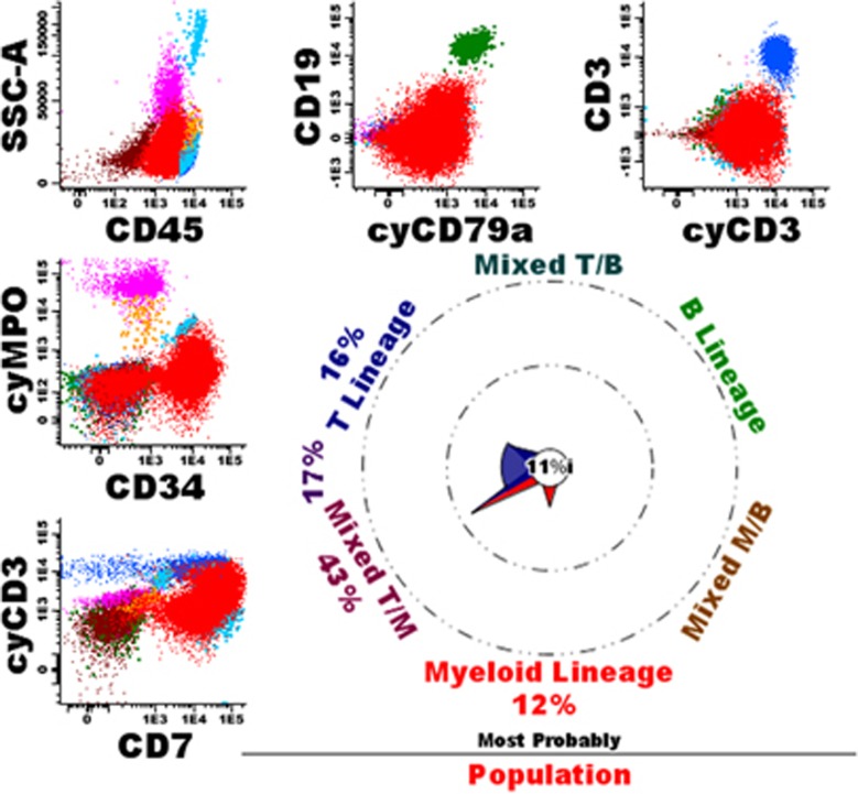
Figure 4.
Example of a patient with an AML with minimal differentiation. The overall immunophenotype was CD34+/CD117+/weak HLADR+/weak CD45+/CD13-/partially CD33+/CD11b-/CD14-/ CD15-/CD16-/partially TdT+/CD36-/CD64-/CD56-/CD4-/heterogeneous CD123+/CD2-/CD5-. Morphology showed an AML, FAB-M0. Cytogenetics were 46,XY, t(1;11)(q21;q23). According to WHO2008 the patient was diagnosed as AML (with minimal differentiation). Normal T-cells (dark blue), NK cells (middle blue), mature B-cells (green), eosinophils (light blue), granulocytes (purple), monocytes (orange) and immature erythroid cells (brown) are shown as well.

