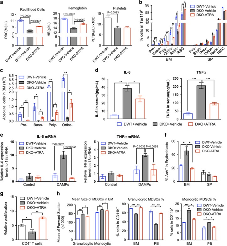
Figure 5.
Amelioration of anemia in DKO mice by targeting MDSCs with AT-RA treatment. (a) DWT or DKO mice were intraperitoneally injected with 400 μg ATRA dissolved in 100 μl DMSO, or equal volume of vehicle control every two days for three doses. 48 h after the last injection, red blood cell counts and hemoglobin levels were assayed. (b, c). The percentages of erythroid cells at different stages were examined by flow cytometry in Ter119-positive cells from the bone marrow and spleen from mice in a. The absolute erythroblasts cell number was calculated in c. (d) Serum TNFa and IL-6 cytokine levels were determined in mice with or without ATRA treatment. (e) Gr1 and Mac1 double-positive MDSCs were purified from the indicated mice and challenged with 1:20 DAMPs for 2 h in vitro. The mRNA levels of TNFa and IL-6 were assayed by real-time PCR using 18 s rRNA as an internal control. (f) The apoptotic erythroblasts in the bone marrow and spleen from the indicated mice were analyzed by Annexin V and PI staining followed by flow cytometric analysis. (g) Gr1 and Mac1 double-positive MDSCs were purified and co-cultured with cell proliferation dye labeled-splenic T cells at 1:1 ratio, the mean fluorescence intensity (MFI) were determined by flow cytometric assay after 48 h stimulation by anti-CD3/CD28 coated Dynabeads. The proliferation rate was calculated as percentage decrease of MFI relative to cells without stimulation. (h) Granulocytic (CD11b+Ly6G+Ly6Clow) and monocytic (CD11b+Ly6G-Ly6high) MDSCs from the bone marrow and peripheral blood of mice in a were assayed by flow cytometry. N=3−5 mice in each group. *P<0.05, **P<0.01, ***P<0.001.

