Figure 1.
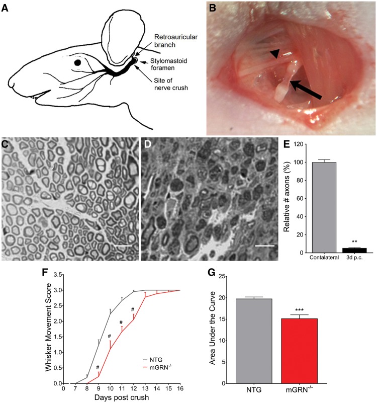
mGRN−/− mice show delayed functional recovery after facial nerve crush injury. (A) Schematic representation of the facial nerve anatomy (adapted and adjusted from Kuzis et al. (55)). The facial nerve crush injury is performed immediately posterior from the bifurcation with the retroauricular branch (B, arrowhead), making the facial nerve completely transparent at the injury site (B, arrow). Semi-thin cross sections of contra- (C) and ipsilateral (D) facial nerve, distal from the injury site. (E) 95% of axons are lost in the distal nerve segment at 3 days post crush. **P < 0.01, Mann-Whitney test. (F) The absence of mGRN significantly delays functional recovery of the of the whisker movement. #P < 0.0001, RM Two-way ANOVA followed by Bonferroni correction. (G) Area under the curve of whisker movement recovery scores is significantly reduced in mGRN−/− mice. ***P < 0.001, Student’s t-test. Data is shown as mean ± SEM.

