Figure 3.
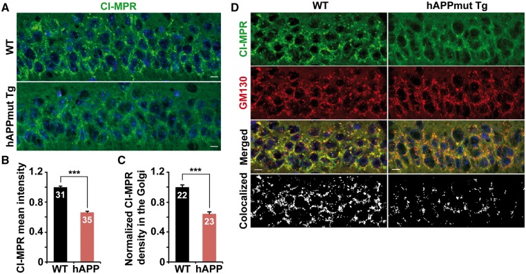
Impaired Golgi targeting of CI-MPR in the soma of mutant hAPP Tg mouse neurons. (A and B) Reduced density of somatic CI-MPR in the hippocampal neurons of mutant hAPP Tg mouse brains. The mean intensity of somatic CI-MPR in hippocampal regions per imaging slice section (320 μm × 320 μm) was quantified. (C and D) Quantitative analysis (C) and representative images (D) showing that CI-MPR trafficking to the Golgi is decreased in hippocampal regions of mutant hAPP Tg mice. The Golgi targeting of CI-MPR was expressed as co-localized intensity of CI-MPR with GM130 (a Golgi marker) in the soma of hippocampal neurons. Scale bars: 10 μm. Data were quantified from a total number of imaging hippocampal slice sections indicated on the top of bars (B and C) from three pairs of mice. Error bars represent SEM. Student’s t test: ***P < 0.001.

