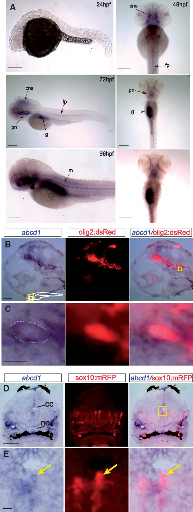
Figure 2.

(A) Bright-field images of whole-mount embryos, in situs for abcd1, ages 24-96 hpf. At 24 hpf no abcd1 expression is seen. By 48 hpf expression is seen in the CNS and floorplate, and from 72-96 hpf additional expression becomes visible in the muscles, pronephros/adrenal glands, and gut. Left panel’s lateral views, dorsal to the top, rostral to the left; right panels, dorsal view, rostral to the top; scale bar 20 µm; cns, central nervous system; fp, floorplate; g, gut; m, muscles; pn, pronephros. (B,C) Section of 72 hpf Tg(olig2:dsRed) embryo double-stained for abcd1 in situ and α-dsRed. abcd1 is expressed in dsRed-labeled oligodendrocyte precursors in the brain. Area shown at higher resolution in C is the region boxed in far-right panel of B. Dotted line in C indicates the approximate boundary of cell membrane. Sagittal view in midline, rostral to the left, dorsal to top, scale bar 5 μm in B; 1 μm in C. (D,E) Cross-section of 72 hpf Tg(sox10:mRFP) embryo double-stained for abcd1 in situ and α-RFP in the spinal cord, dorsal to the top. abcd1 is expressed in dedicated oligodendrocyte precursors (arrow). Scale bar 10 μm in D, 1 μm in E; abbreviations: cc, central canal; nc, notochord.
