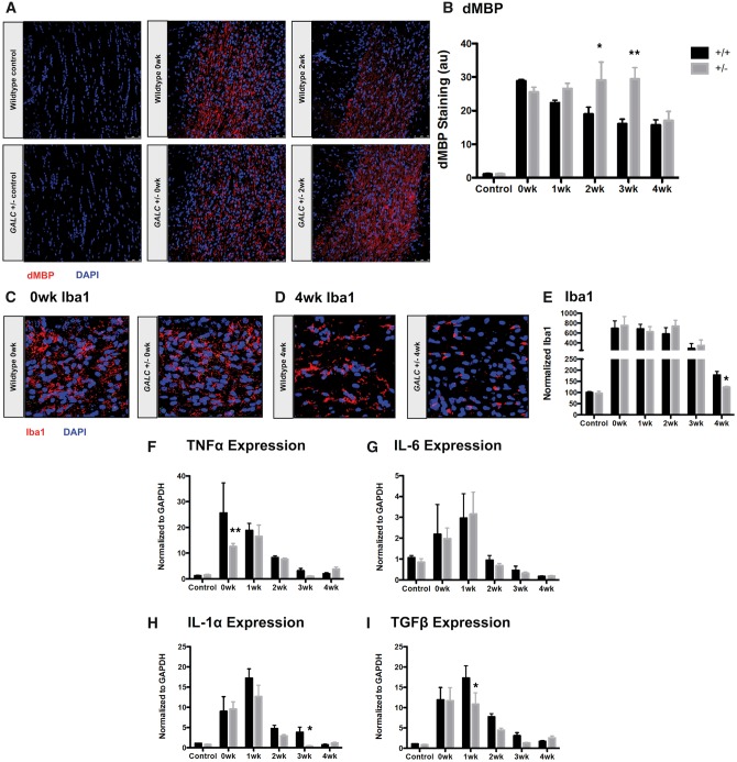
Figure 3.
GALC +/− animals have elevated damaged myelin and altered microglial response following cuprizone exposure. (A,B) Representative confocal images and quantification of damaged Myelin Basic Protein (dMBP) in the corpus callosa of three-month-old WT and GALC +/− littermates after 4 weeks exposure to 0.3% cuprizone diet (N = 3 from different litters per time point). (C–E) Representative confocal images and quantification of Iba-1 microglial staining in (C) 0-week cuprizone recovery WT and GALC +/− animals, (D) 4-week cuprizone recovery WT and GALC +/− animals, and (E) quantification of Iba-1 staining in the corpus callosa of three month old WT and GALC +/− littermates after 4 weeks exposure to 0.3% cuprizone diet (N = 3 from different litters per time point). Data are normalized to WT controls. (F–I) Quantification of the relative levels of mRNA of (F) tumor necrosis factor alpha (TNFα), (G) interleukin-6 (IL-6), (H) interleukin-1 alpha (IL-1α), and (I) transforming growth factor beta (TGFβ). Data are normalized to GAPDH and represented as fold change over WT untreated controls (N = 3 from different litters). Data for all graphs displayed as mean ± SEM. *P < 0.05, **P < 0.01 versus WT recovery-matched animals.

