Figure 3.
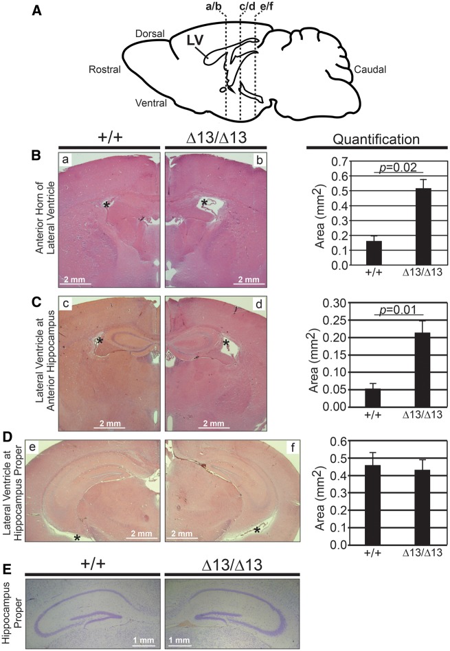
Analysis of brain and hippocampal morphology in Zc3h14Δex13/Δex13 mice. (A) Diagram of lateral view of mouse brain and location of coronal sections shown in the following histological images. LV, lateral ventricle. Representative H&E stains of lateral ventricles of Zc3h14+/+ (+/+) and Zc3h14Δex13/Δex13 (Δ13/Δ13) adult mice at the following level of slices: (B) anterior horns of ventricles (a/b in diagram), (C) anterior hippocampus (c/d in diagram), and (D) hippocampus proper (e/f in diagram). Magnification ×2. Scale bars, 2 mm. *, lateral ventricle. Shown to the right are the corresponding quantifications of the area (mm2) of the lateral ventricles at the indicated levels for Zc3h14+/+ and Zc3h14Δex13/Δex13mice. Error bars indicate SEM. For quantification of anterior horns of lateral ventricles; Zc3h14+/+, n = 3; Zc3h14Δex13/Δex13, n = 5. For quantification of lateral ventricles at anterior hippocampus; Zc3h14+/+, n = 3; Zc3h14Δex13/Δex13, n = 3. For quantification of lateral ventricles at hippocampus proper; Zc3h14+/+, n = 6; Zc3h14Δex13/Δex13, n = 6. (E) Comparable brain sections from Zc3h14+/+ (+/+) and Zc3h14Δex13/Δex13 (Δ13/Δ13) adult mice were stained with cresyl violet. Scale bars equal 1 mm.

