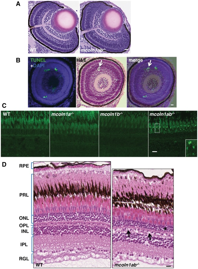
Figure 5.
Eye pathology in mcoln1 mutant zebrafish at both embryonic and adult stages. (A) H&E staining on paraffin sections of 3 dpf WT and mcoln1ab−/− zebrafish embryos showing the eye structure. Scale bar, 20 μm. (B) TUNEL and DAPI co-staining on a 3dpf eye paraffin section of mcoln1ab−/− zebrafish is merged together with an H&E stained section that is 20 μm apart, the white arrow shows a TUNEL positive apoptotic body-like structure. Scale bar, 20 μm. (C) Auto-fluorescence of 8-month-old zebrafish retina on cryostat sections for WT, mcoln1a−/−, mcoln1b−/−, and mcoln1ab−/−. Insets show a twofold magnification of the indicated region. Scale bar, 5 μm. (D) H&E staining on paraffin sections of 8-month-old WT and mcoln1ab−/− zebrafish retina showing the structure of different retinal layers. The black asterisk indicates thinning of the ONL in mcoln1ab−/− zebrafish. The black arrows mark cell loss in the INL. RPE, Retinal Pigment Epithelium; PRL, Photoreceptor Layer; ONL, Outer Nuclear Layer; OPL, Outer Plexiform Layer; INL, Inner Nuclear Layer; IPL, Inner Plexiform Layer; RGL, Retinal Ganglion Layer. Scale bar, 10 μm. All the images are representative of at least three independent experiments.

