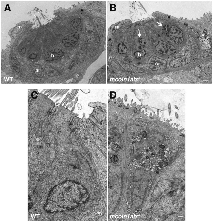
Figure 8.
Ultrastructure of hair cell in mcoln1ab−/− larvae at 5 dpf. Transmission Electron Microscopy (TEM) for one neuromast in both WT (A) and mcoln1ab−/− (B) larvae at 5 dpf. m stands for mantle cell, h stands for hair cell, and s stands for support cell. White arrows mark the inclusion bodies in mcoln1ab−/− hair cells, and the asterisk marks inclusion bodies surrounding the hair cells. Scale bar, 1 μm. (C) and (D) Higher magnification showing more details of one of the hair cells in both WT (C) and mcoln1ab−/− (D). Scale bar, 200 nm.

