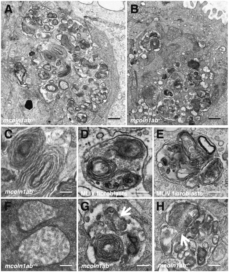
Figure 9.
Autophagic dysfunction in mcoln1ab−/− hair cells at 5 dpf. (A,B) Higher magnification of TEM showing autophagic vacuoles containing electro-dense inclusions, organelles and other cytoplasmic contents in mcoln1ab−/− hair cells. (C) Typical lamellar structure in mcoln1ab−/− hair cells. (D,E) Typical lamellar structures in MLIV fibroblasts. (F) Electron-transparent matrix and reduced cristae in mcoln1ab−/− hair cell mitochondria. (G,H) Autolysosomes containing partially degraded mitochondria inside in mcoln1ab−/− hair cells. Scale bar for (A) and (B), 200 nm. Scale bar for (C) to (H), 80 nm.

