Figure 1.
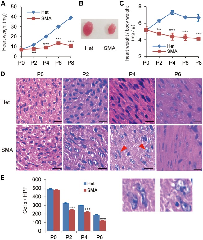
Heart weight and cardiomyocyte density of SMA mice are dramatically reduced, compared with heterozygotes. (A) Comparison of heart weight between SMA mice and their heterozygous littermates (Het) at five time points (P0, P2, P4, P6, and P8) during postnatal development (n = 8). (B) Representative pictures of hearts obtained from P4 SMA and heterozygous mice. (C) Comparison of the heart weight/body weight ratios from P0 to P8 between SMA and heterozygous mice (n = 8). (D) Hematoxylin and eosin (H&E) staining of sections from hearts of heterozygous (upper) and SMA (lower) mice on P0, P2, P4, and P6. The two enlargements below show irregular nuclei of cardiomyocytes (P4, SMA). Scale bar, 50 μm. (E) Quantitation of the number of cardiomyocytes per high-power field (HPF) (n = 4). **P < 0.01, ***P < 0.001.

