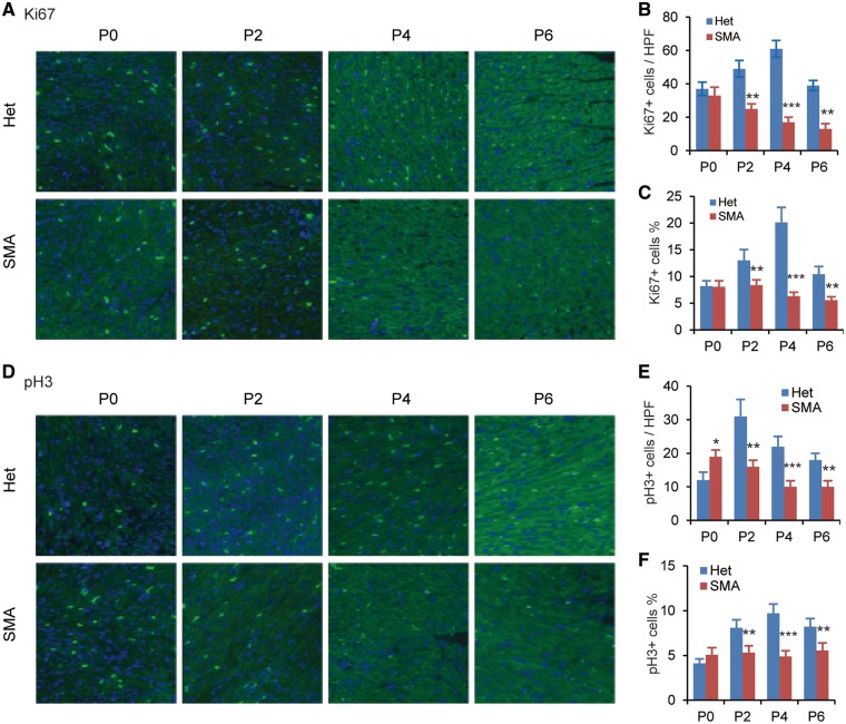
Figure 2.
Cardiomyocyte proliferation and cell cycle are impaired in SMA mice. Heart sections from SMA (n = 3) and heterozygous (Het, n = 3) mice at four time points (P0, P2, P4, and P6) were stained with anti-Ki67 antibody (A) or anti-pH3 antibody (D) to label mitotic cells; nuclei were counterstained with DAPI. Total number of Ki67+ (B) or pH3+ cardiomyocytes (E) and percentage of Ki67+ (C) or pH3+ (F) cardiomyocytes per high-power field (HPF) was determined from three sections per heart, with three hearts per group (n = 9). **P < 0.01, ***P < 0.001.

