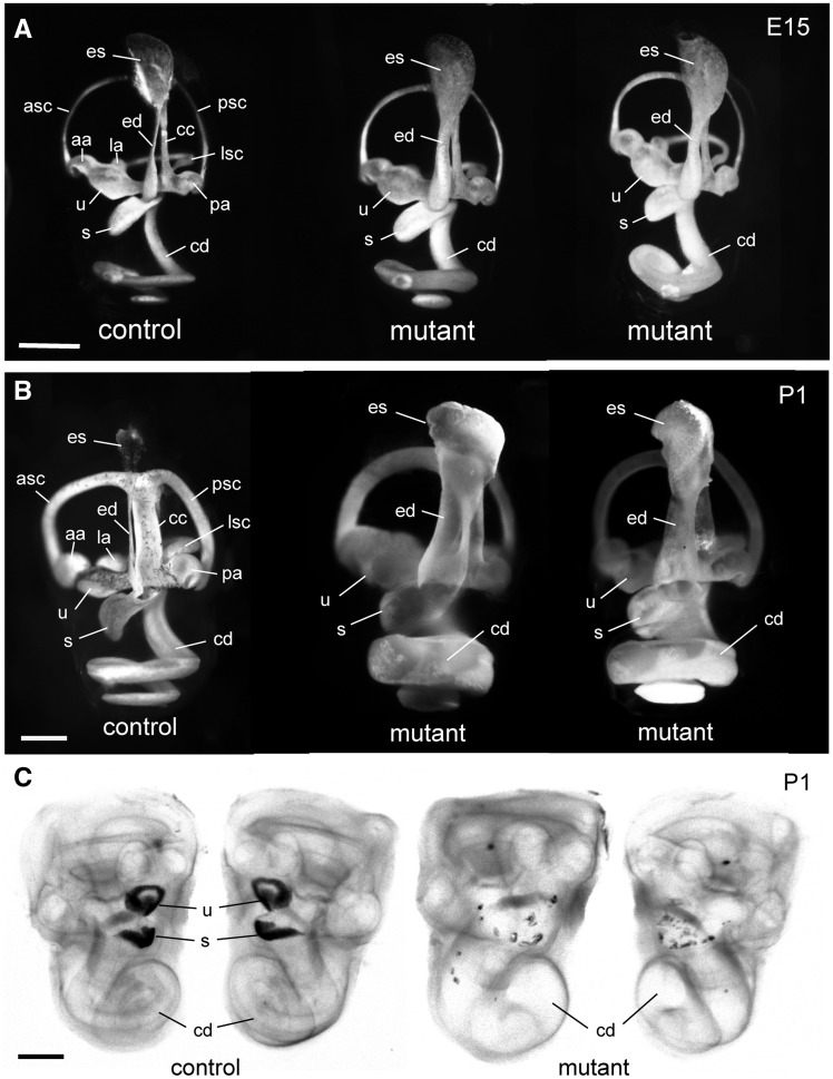
Figure 3.
Paint fills and whole mounts of inner ears from MRL-Atp6v1b1vtx/vtxmutant and control mice. (A) Paint fills of the membranous labyrinths of inner ears from an E15-stage MRL-Atp6v1b1+/vtx (control) embryo and two littermate MRL-Atp6v1b1vtx/vtx (mutant) embryos. The endolymphatic sac (es), endolymphatic duct (ed), utricle (u), saccule (s), and cochlear duct (cd) appear enlarged in the two mutant inner ears compared with the control. (B) Paint fills of the membranous labyrinths of inner ears from a newborn (P1) MRL-Atp6v1b1+/vtx (control) mouse and two MRL-Atp6v1b1vtx/vtx (mutant) littermates. The entire membranous labyrinth of the mutant inner ears appears swollen from an excess of endolymph. Other structures labeled in the control inner ears of A and B: anterior semicircular canal (asc), posterior semicircular canal (psc), lateral semicircular canal (lsc), anterior ampulla (aa), posterior ampulla (pa), lateral ampulla (la), common crus (cc). (C) Cleared, whole-mount preparations of both inner ears from a P1 Atp6v1b1+/vtx newborn mouse (control) and two MRL-Atp6v1b1vtx/vtx (mutant) littermates, illuminated from below. Dark-appearing otoconia in the utricle (u) and saccule (s) are clearly visible in the control ear, but only a few widely dispersed otoconial crystals are seen in the mutant inner ears. The cochlear ducts (cd) of the mutant inner ears are enlarged compared with the control. Scale bars for A, B, C: 0.5 mm.

