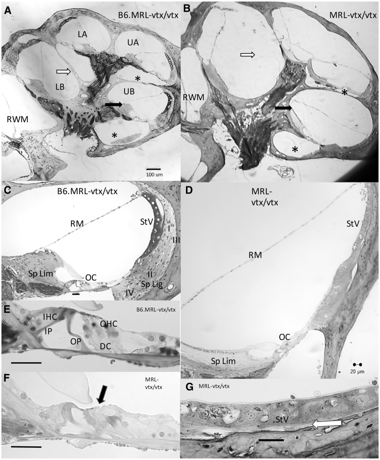
Figure 4.
Cross sections of cochleae from B6.MRL-Atp6v1b1vtx/vtx and MRL-Atp6v1b1vtx/vtx mice. (A, B) Whole cochlear profiles from B6.MRL-Atp6v1b1vtx/vtx (A) and MRL-Atp6v1b1vtx/vtx mice (B) imaged at the same magnification. B6.MRL-Atp6v1b1vtx/vtx mutants appear normal while MRL-Atp6v1b1vtx/vtx mutants feature greatly reduced scala tympani (compare areas marked with *), missing interscalar septum between lower base and lower apex (compare areas marked with white arrows), and differences in the shape of spiral limbus and attachment point of Reissner’s membrane (compare black arrows). (C, D) Upper basal turn scala media profiles from different specimens of B6.MRL-Atp6v1b1vtx/vtx (C) and MRL-Atp6v1b1vtx/vtx mice (D) imaged at the same magnification. B6.MRL-Atp6v1b1vtx/vtx mutants appear normal while MRL-Atp6v1b1vtx/vtx mutants show widened profile, elongated and acellular spiral limbus, hair cell loss (see E, F), and spiral ligament degeneration (see G). (E, F) Enlarged views of organ of Corti from C,D show outer hair cell loss in MRL-Atp6v1b1vtx/vtx mutant (black arrows). (G) Enlarged view of MRL-Atp6v1b1vtx/vtx mutant stria vascularis from D shows thin and somewhat disorganized stria with near absence of type I and III fibrocytes (white arrow). Scale bar in A applies to B. All other scale bars 20 µm. LB, Lower base; UB, Upper base; LA, Lower apex; UA, Upper apex; RWM, Round window membrane; Sp Lim, Spiral Limbus; Sp Lig, Spiral ligament; RM, Reissner’s membrane; StV, Stria vascularis; I, II, III, IV, Fibrocyte types by area; OC, Organ of Corti; IP, Inner pillar; OP, Outer pillar; DC, Deiters’ cells; IHC, Inner hair cell; OHC, Outer hair cell.

