Figure 3.
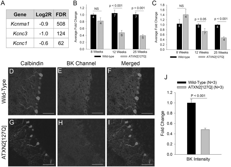
A progressive reduction in potassium channel transcripts accompanies degeneration in ATXN2[127Q] Purkinje neurons. (A) RNA sequencing from 6-week-old cerebella of ATXN2[127Q] mice and littermate controls reveals a reduction in transcripts of potassium channels important for Purkinje neuron spiking. FDR (false discovery rate) >30 corresponds to a corrected p value of < 0.01. (B) Quantitative RT-PCR for Kcnma1 (BK) demonstrates a progressive reduction in BK channel transcripts in ATXN2[127Q] cerebella. (C) Quantitative RT-PCR for Kcnc3 (Kv3.3) demonstrates a progressive reduction in Kv3.3 channel transcripts in ATXN2[127Q] cerebella. Immunostaining for calbindin (D) and BK channels (E) in wild-type Purkinje neurons shows prominent overlap of BK and calbindin staining (F). (G) In 25-week-old ATXN2[127Q] cerebella, calbindin immunostaining reveals prominent Purkinje neuron dendritic atrophy, with thinning of the molecular layer. (H) BK staining is reduced in ATXN2[127Q] Purkinje neurons, also seen in the merged image of BK and calbindin in (I) and summarized in (J). Data were analysed with a Student’s t-test, and are displayed as mean ± SEM.

