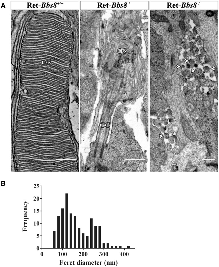
Figure 6.
Aberrant photoreceptor ultrastructure in Bbs8-null mice. (A) Left: TEM images of photoreceptor OSs in P10 retinas of wild-type (left; scale bar, 500 nm) and Bbs8-null (center panel; scale bar, 500 nm) mice. In contrast to the well-ordered disc stack seen in wild-type controls, knockout animals possessed shortened and dysmorphic OSs, with highly disorganized disc membranes. Retinas from knockout animals also displayed 100-300 nm extracellular vesicles (arrowheads), commonly observed adjacent to the proximal ISs (right panel; scale bar, 500 nm); extracellular vesicles were never observed in the wild-type controls. (B) Distribution of Feret diameter measurements (nm) of extracellular vesicles found in Bbs8−/− animals at P10 (N = 142 from two animals).

