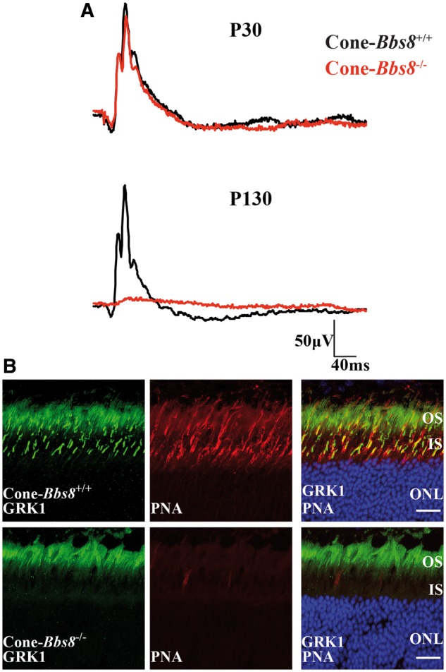
Figure 8.

Degeneration of cone photoreceptors missing BBS8. (A) Representative trace of photopic ERG responses measured under light adapted conditions at 0.69 cd.s−1m2 in Cone-Bbs8−/− and littermate controls at P30 and P130. (B) Retinal cryosections stained against G-protein-coupled Receptor Kinase 1 (GRK1; green) and Peanut agglutinin (PNA) in Ret-Bbs8−/− and wild type littermate controls at P130. GRK1 stains both rods and cones while PNA marks the extracellular matrix surrounding the cones. Scale bar: 20 μm.
