Figure 3.
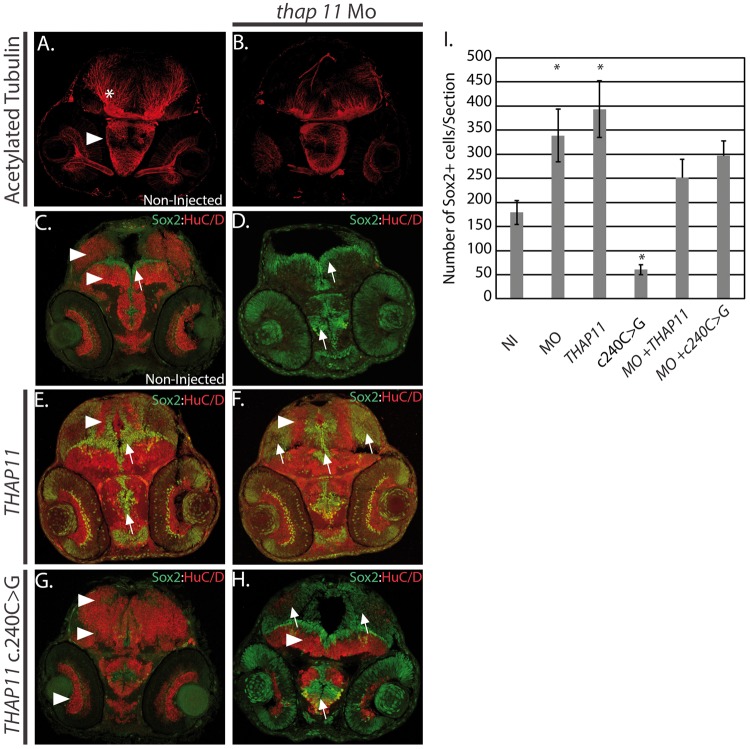
Thap11 regulates neural stem cell proliferation and differentiation. (A,B) 2 day post fertilization (dpf) zebrafish embryos analysed for anti-acetylated tubulin expression using antibody immunohistochemistry. Arrowhead indicates hypothalamus and asterisk labels the presumptive cerebellum. (A) Non-injected (NI), (B) thap11 morpholino (thap11 Mo). (C–H) Immunohistochemistry with anti-Sox2 (green) and anti-HuC/D (red) antibodies at 2 day post fertilization (dpf) from (C) non-injected, (D) thap11 morphants (thap11 Mo), (E) THAP11 mRNA injected (THAP11), (F) thap11 morphants co-injected with THAP11 mRNA, (G)THAP11 c. 240C>G mRNA injected, and (H) thap11 morphants co-injected with the THAP11 c240C>G. Arrowheads demonstrate regions of HuC/D expression and arrows indicate Sox2 expression. HuC/D expression is consistently observed in the optic tectum and tegmentum, regions indicated by the arrowheads in (C). A thin lining of neural precursors are present surrounding the ventricle region and indicated by the arrow. Anatomically comparable regions from serial sections are demonstrated in the treated groups. n = 9 per group. I. Bar graph showing the cell counts of Sox2+ cells in sections from C-H. NI= non-injected, MO = thap11 morpholino injected, THAP11 = wild type THAP11 mRNA injected, c240C>G = mutant THAP11 mRNA injected, MO + THAP11 = co-injected as in (F), MO + c240C>G = co-injected as in (H). The counts were carried out in 9 sections in each category (n = 9) and error bars are shown. Asterisks denote results that were statistically significant.

