Figure 1.
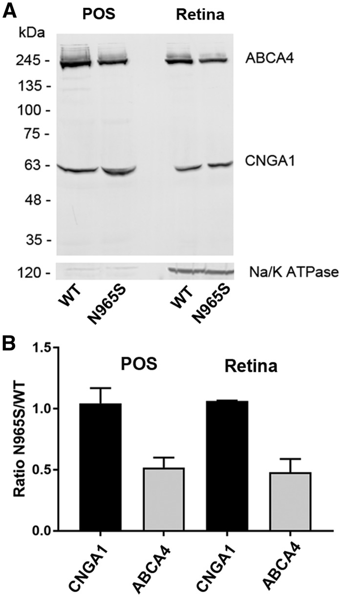
Quantification of ABCA4 in WT and p.Asn965Ser photoreceptor outer segments and retina membranes by Western blotting. Enriched photoreceptor outer segments (POS) and retinal membranes (Retina) were prepared from WT and p.Asn965Ser ABCA4 mice. (A) Equal amounts of protein from POS (20 µg) and Retina (50 µg) preparations isolated from WT and homozygous p.Asn965Ser (N965S) mice were applied to the lanes of a SDS polyacrylamide gel and resulting western blots were labeled with the Rim3F4 antibody to ABCA4, the PMc1D1 antibody to the CNGA1 channel, and an antibody to the Na/K ATPase (α subunit) as a marker for inner segments. (B) Quantification of ABCA4 and CNGA1 in POS and Retina from p.Asn965Ser and WT mice. The amount of CNGA1 in the p.Asn965Ser preparations was identical to the amount in WT preparations, whereas quantity of p.Asn965Ser (N965S) ABCA4 was about half that of WT ABCA4 in both POS and Retina preparations. Data are presented as the mean ± SD for three independent preparations.

