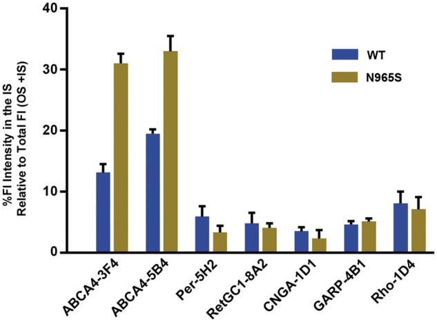
Figure 4.

Percent immunofluorescence staining intensity of ABCA4 and other outer segment proteins in the inner segment (IS). Data obtained from immunofluorescence microscopy of WT and p.Asn965Ser (N965S) cryosections as per Figures 2 and 3. Ten squares (6 × 6 µm) were selected in each the inner and outer segment layers and the mean fluorescence pixel intensities were obtained using Image J software. The data are expressed as the % unit area of fluorescence (Fl) intensity in the IS relative to the total intensity (IS + OS). The data provide a quantitative assessment of the staining of antibodies in the inner vs. the outer segment. ABCA4 shows significant staining in the IS for WT and p.Asn965Ser (N965S) photoreceptors with increased staining in the IS for the p.Asn965Ser photoreceptors relative to WT photoreceptors. Data from two sets of mice were similar.
