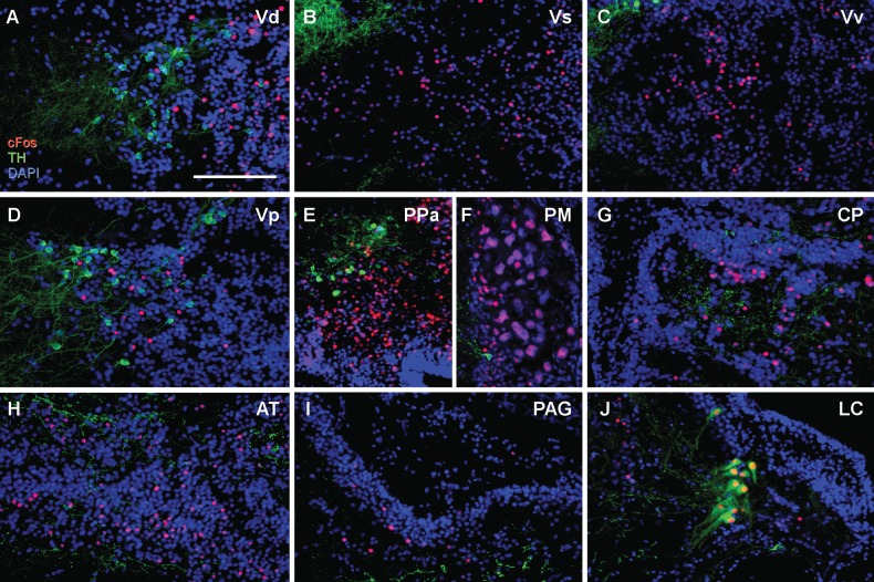
Fig. 2.
Example micrographs showing nuclei found to have no significant relationship between time attending speaker and TH-ir + cFos colocalization (A,D,E,J) or cFos expression (A–I). Transverse sections are shown from the ventral telencephalon (V) (A-D): (A) dorsal nucleus of V (Vd), (B) supracommissural nucleus of V (Vs), (C) ventral nucleus of area V (Vv), and (D) postcommissural nucleus of area V (Vp). Diencephalon (E–H): (E) anterior parvocellular preoptic nucleus (PPa), (F) magnocellular preoptic nucleus (PM), (G) central posterior nucleus of the thalamus (CP), (H) anterior tuberal nucleus (AT). Brainstem (I–J): (I) periaqueductal gray (PAG), (J) locus coeruleus (LC). Scale bar = 100 µm.

