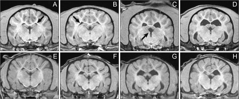
Figure 5.
Examples of small (A), medium (B), large (C), and very-large (D) lateral brain ventricles in the adult dogs (top) and juvenile dogs (bottom) at baseline MRI images. No very large ventricles were found in the juvenile group based on adult dog experience, so only small (E), medium (F), and large (G) juvenile lateral ventricles are shown along with a small right and a large left lateral ventricle to demonstrate an example of asymmetry seen in this study (H). While G of the juvenile dogs appears similar to D of the adult dogs, baseline assessment was based on 3D visualization, whereas the images in this 2D figure were chosen to show the ventricles at a similar cross section of all animals. Qualitative assessment of the ventricle size and distribution of differently sized ventricles between groups was warranted as not to skew group means for the adult and juvenile dogs’ treatment groups. Arrows point to the left lateral, right lateral, and third ventricle in A, B, and C, respectively.
[Reprinted from Hines CDG, Song X, Kuruvilla S, Farris G, Markgraf CG. 2015. Magnetic resonance imaging assessment of the ventricular system in the brains of adult and juvenile beagle dogs treated with posaconazole IV Solution, J Pharmacol Toxicol Methods, with permission from Elsevier.]

