
Figure 6. Decreased oxidative damage and increased anti-oxidant capacity after juvenile MCAo compared to adult.
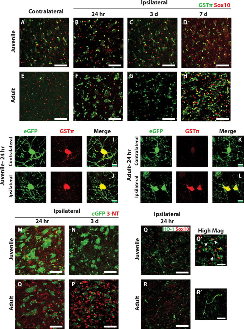
(A–H) Ischemic and control juvenile (A–D) and adult (E–H) sections were immunostained to detect GSTp (green) and Sox10 (red). High basal expression of GSTp was observed in control juvenile striatal oligodendrocytes (A) and low basal expression in control adult striatal oligodendrocytes (E). GSTp expression remained high in juvenile striatal oligodendrocytes after MCAo (B–D) and was upregulated by remaining adult oligodendrocytes after MCAo (F–H). (I–L) High magnification images of PLP-EGFP-expressing oligodendrocytes 24 h after MCAo demonstrate high GSTp expression in EGFP-expressing oligodendrocytes in both the control contralateral (I) and injured ipsilateral (J) striatum of juvenile mice. Low GSTp expression was observed in control contralateral EGFP-expressing oligodendrocytes in adult mice (K) and was upregulated in injured EGFP-expressing oligodendrocytes in the ipsilateral striatum (L). Ischemic PLP-EGFP-expressing juvenile (M, N) and adult (O, P) sections were immunostained to detect nitrotyrosine (3-NT, red). Moderate 3-NT expression was observed in juvenile animals at 24 hr after MCAo (M), which generally resolved 3 days after MCAo (N). High 3-NT expression was observed in adult animals at 24 hr post-MCAo (O) and it remained high after 3 days (P). Sections were also immunostained to detect HO-1 (Q, R, green) and Sox10 (red). High HO-1 was observed in juvenile sections at 24 hr post-MCAo (Q), and often colocalized with Sox10 expressing oligodendrocytes (Q’, arrowheads). Adult expression of HO-1 after MCAo was much lower (R). White scale bars: 100 lm (A–H, M–R), 40 lm (Q’, R’). Green scale bars: 10 lm (I–L).