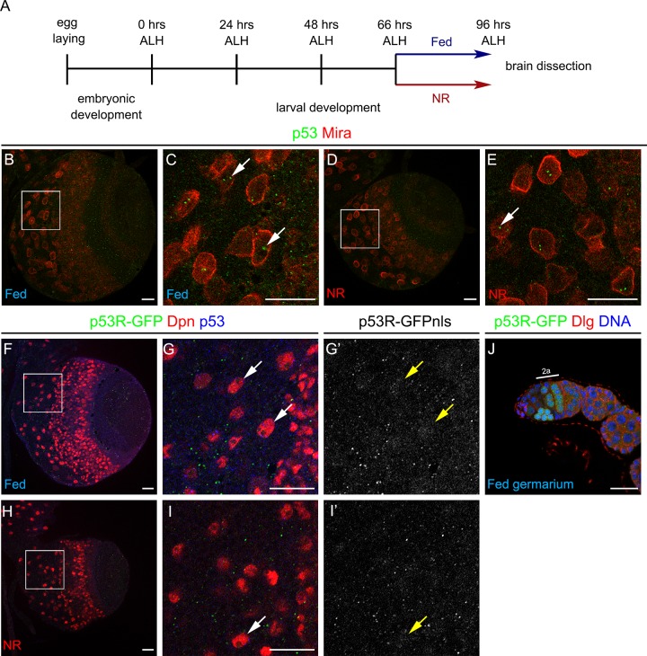
Fig 3. Drosophila p53 levels and activity in neural stem cells are independent of the nutritional condition.
(A) Schematic representation of the nutrient restriction (NR) protocol. (B-E) Immunostaining against p53 (green) and Miranda (Mira, in red) of wild-type (w1118/1118) larval brain under (B, C) Fed or (D, E) NR conditions. (F-I’) Staining against GFP (green and gray), Deadpan (Dpn, neural stem cell marker in red) and p53 (blue) of larval brains of p53 reporter (p53R-GFP) under (F-G’) Fed or (H-I’) NR conditions. Arrows point to neural stem cells. (J) Ovariole of p53 reporter stock (p53R-GFP) stained for GFP (green), Dlg (red) and DNA (blue). Scale bars are 20 μm.

