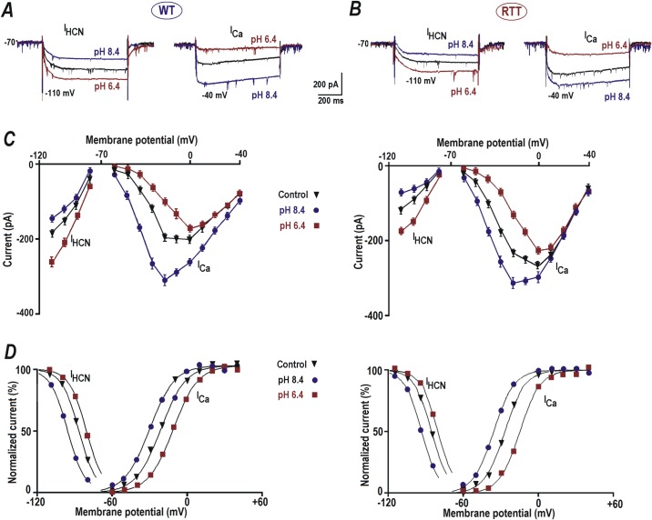
Fig 2. Modulation of HCN and calcium currents by extracellular pH.
A and B–Sample HCN and VSCC currents from WT and RTT respectively. The currents were evoked by voltage steps to -110 and -40 mV, respectively, from a holding potential of -70 mV (close to resting potential measured in CA1 neurons). The cells were patched using Cs+ + TEA intracellular solution (see Methods) that allowed clear isolation of the hyperpolarization-activated (IHCN) and depolarization-evoked calcium (ICa) currents; which were activated below -70 and above -60 mV, respectively. HCN current was smaller and VSCC current was bigger in RTT CA1 neurons in comparison to WT (A and B, black traces). A decrease in pH to 6.4 increased the HCN current (A and B left, brown traces) and reduced the Ca2+ current (A and B right, brown traces). pH increase to 8.4 caused a reduction in HCN current (A and B left, blue traces) and potentiation (A and B right, blue traces) of the calcium current. C and D—I-V curves for steady state HCN and VSCC currents from WT and RTT CA1 cells. E and F—Activation curves (m∞) determined from the tail currents. The points represent the means measured in four CA1 cells from WT and RTT slices. The data were mean-square fitted with the Boltzmann function m∞ = 1/ [1 + exp (-(V—V1/2)/b)]. The slope factor of b = 8 mV was found to be the same for HCN and VSCC currents.

