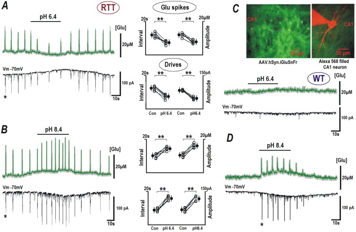
Fig 3. Modulation of glutamate spikes and bursting activity by extracellular pH.
RTT and WT slices were transduced with neuron-targeted virus with glutamate sensor iGluSnFr (see Materials and methods). Upper traces in each panel indicate mean glutamate changes (obtained from 12 neurons in the image field) overlaid upon grey background indicating ± SEM. Brief glutamate transients in RTT slices appeared synchronously in the image field. They temporally coincided with the synaptic drives (a correlate of the bursting activity) in the patched CA1 cell (lower block traces in each panel). ACSF with pH 6.4 suppressed the generation of glutamate spikes and the synaptic currents (A). Elevation of pH to 8.4 increased the frequency and amplitude of glutamate spikes and corresponding synaptic currents (B). Group summary for experiments in A and B is presented on the right. The data for RTT were evaluated before and after pH effects stabilized (ca. 2 min), with Student’s t test with confidence levels of P < 0.01 (**). In the case of WT, neither glutamate levels nor synaptic activity were modified at acidic pH (6.4, C), but during the application of alkaline ACSF with pH 8.4, glutamate spikes and synaptic drives appeared (D). The inset in the right upper corner shows the sensor fluorescence in CA1 area and the image of patched neuron filled with 100 μM Alexa 568 (red) overlaid on the image of neuronal glutamate sensor in the slice (green).

