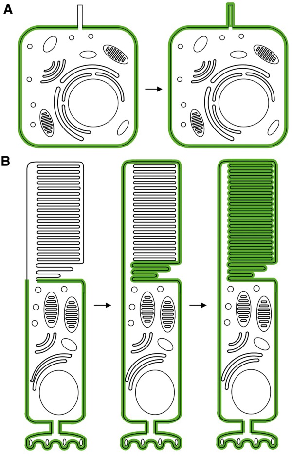
Figure 2.

Photoreceptor outer segments as a membrane protein sink. (A) Localization of a hypothetical membrane protein (green) with no targeting signals in typical cells with primary cilia. Left; when the ciliary gate is intact, right; when the ciliary gate is defective or absent. (B) Localization of a hypothetical membrane protein (green) with no targeting signals in rod photoreceptors. Left; when the ciliary gate is intact and the protein is actively excluded from the OS, middle; soon after the ciliary gate is disrupted, right; after the entire outer segment is renewed. Schematic cells are not drawn to scale.
