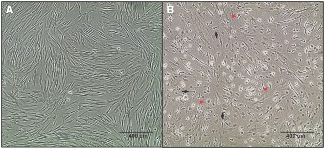
Figure 2.
Morphological changes during neuronal differentiation of DPSC. (A) Undifferentiated DPSC have a spindle-shape similar to fibroblasts. (B) DPSC were differentiated and allowed to mature for 3 weeks, resulting in a mixed culture containing both neuron-like and glial-like cells. The neurons (black arrows) show a pyramidal-like, neuronal morphology with shorter projections similar to dendrites and a longer axonal projection on the opposing side. The glial cells (red arrows) show a typical star-shaped morphology with a rounded cell body and small projections extending from the perimeter. Both images were taken at a 10x magnification using phase-contrast microscopy.

