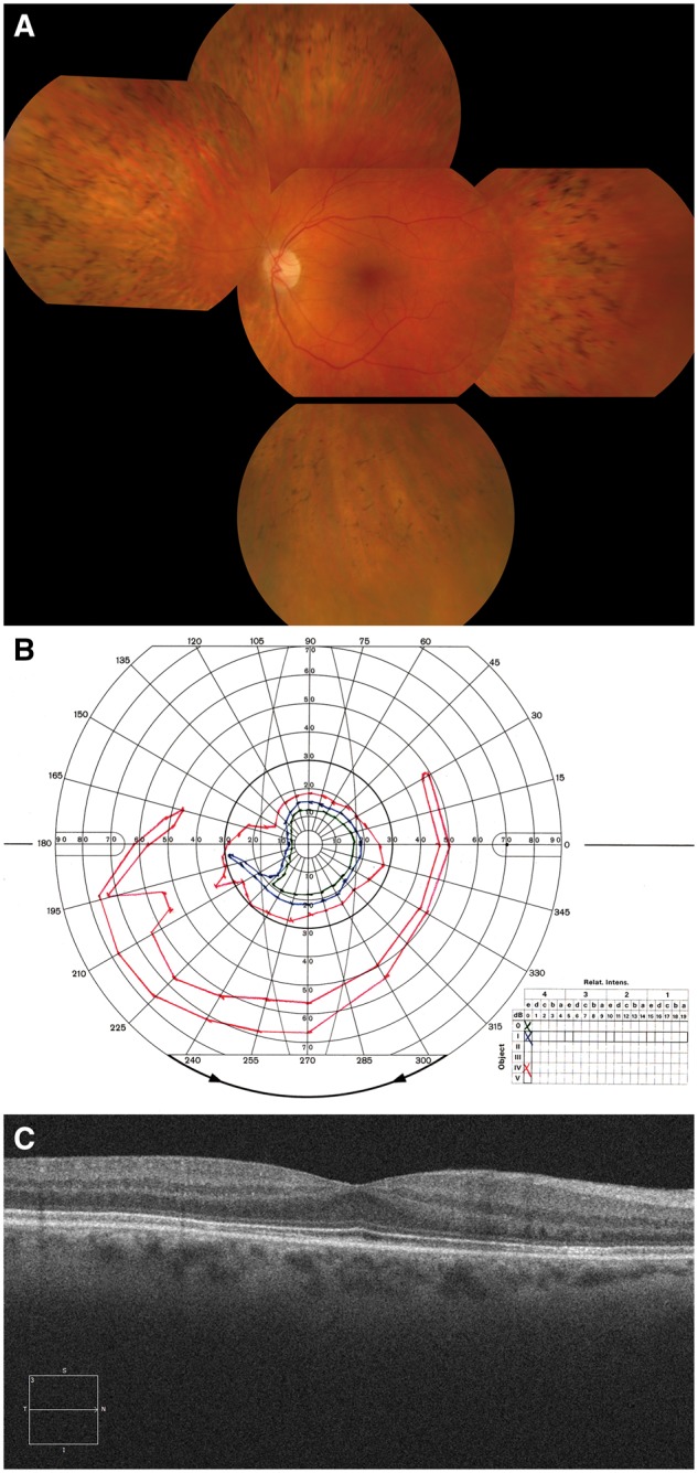
Figure 1.

SLC24A1 causative of an autosomal recessive retinal degeneration. Panel A: Fundus photograph of the right eye of the proband with an SLC24A1 mutation indicating mild optic disc pallor, moderate arteriolar attenuation and quite extensive thinning of the retinal pigment epithelium in the retinal mid-periphery with abundant bone spicule intra-retinal pigment deposits, typically seen in patients with Retinitis Pigmentosa. Panel B: Goldmann kinetic perimetry of the left eye. Marked concentric constriction is evident, even to the large IV4e target (red), with preservation of an inferior island of field. This pattern of visual field loss is very typical of that seen in Retinitis Pigmentosa. Panel C: Optical Coherence Tomography (OCT) image of the left macula showing preservation of retinal structures, consistent with the normal appearance of the macula in Panel A and the patient’s good best-corrected visual acuity of 6/6.
