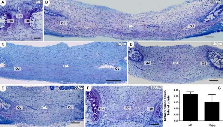
Fig 1. Dynamic changes in the histoarchitecture and ECM deposition accompany PS remodelling.
(A-F) Giemsa staining of PS and IpL transverse sections. (A) NP PS consists of a narrow fibrocartilaginous disc (FC) situated between two high metachromatic hyaline cartilaginous pads (HC) on the surface of the subchondral pubic bones, which are caudally and ventrally connected by non-metachromatic dense connective tissue. (B-E) The absence of highly metachromatic hyaline cartilage (HC) and the presence of IpL attached to the pubic bone via an osteoligamentous junction (OJ). (F) The return of similar NP PS histoarchitecture and highly metachromatic hyaline cartilage (HC) tissue at 10dpp in the PS. (G) Morphometric measurement of hyaline cartilage metachromatic tissues volume in the NP and 10dpp mice PS (U = 3; p = 0.7). Data from a Mann-Whitney test are presented with the means and SEM. (A, F) Scale bars = 50 μm. (B-E) Scale bars = 100 μm.

