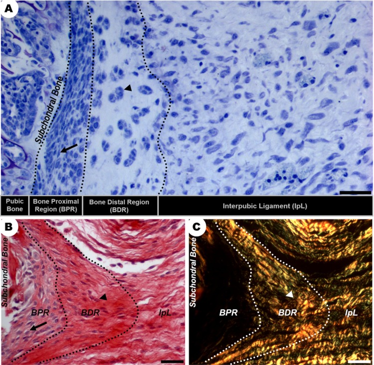
Fig 2. Morphological characterization of distinct regions of the IpL osteoligamentous junction at D19.
(A-C) There are two distinct regions in the IpL osteoligamentous junction (dotted line): the BPR is composed of elongated cells (arrows) with poorly birefringent collagen fibrils at the ECM, while the BDR contains angular chondrocyte-like cells (arrowheads) organized as isogenous groups surrounded by dense bundles of collagen. (A) Giemsa staining, scale bar = 30 μm. (B, C) Sirius Red staining and polarized microscopy, scale bar = 20 μm.

