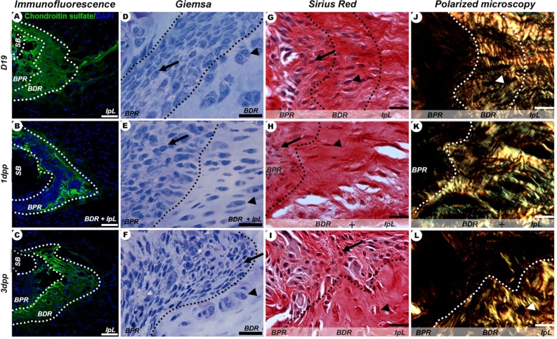
Fig 3. IpL osteoligamentous junction remodelling during late pregnancy (D19) and early postpartum (1-3dpp).
(A-C) Identification of chondroitin sulphate at BDR and BPR regions (dotted lines). (D-I) Typical cell phenotypes present in BPR (arrow) and BDR (arrowhead). (J-L) Isogenous groups of chondrocytes seen as dark areas (arrowhead) distributed in BDR and BPR regions that exhibit differently birefringent collagenous ECM. Scale bar = 20 μm.

