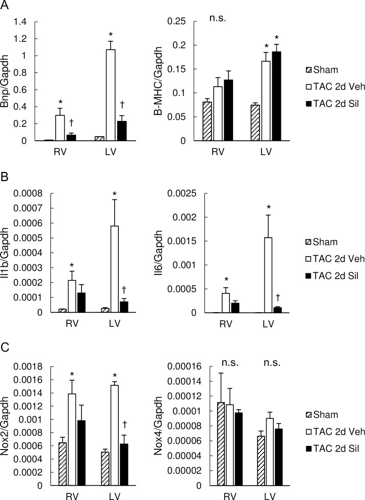
Fig 3. Pathological gene induction in RV as well as LV were ameliorated by sildenafil.
(A) Expression of fetal genes as markers of cardiac hypertrophy, encoding for BNP and B-MHC, normalized to GAPDH. Transverse aortic constriction (TAC) for two days induced marked increase in BNP mRNA levels in the RV myocardium as well as in the LV myocardium, which was prevented by sildenafil. Expression of B-MHC was increased by two-day TAC in LV, but was not affected by sildenafil. (B) Expression of inflammatory cytokines, IL1b and IL6, normalized to GAPDH. IL1b and IL6 was up-regulated by TAC in both ventricles. Sildenafil inhibited both in LV, and also attenuated both in RV. (C) Expression of genes for enzymes inducing oxidative stress, NOX2 and NOX4, normalized to GAPDH. Expression of NOX2 was increased by TAC in both ventricles, and suppressed by sildenafil in LV. TAC and sildenafil had little influence on NOX4 expression. Results are expressed as mean ± s.e.m. (n = 5). TAC 2d Veh, TAC for 2 days with vehicle treatment; TAC 2d Sil, TAC for 2days with sildenafil treatment. n.s., not significant by one-way analysis of variance; *, p<0.05 versus sham group; ✝, p<0.05 versus the TAC 2d Veh group.

