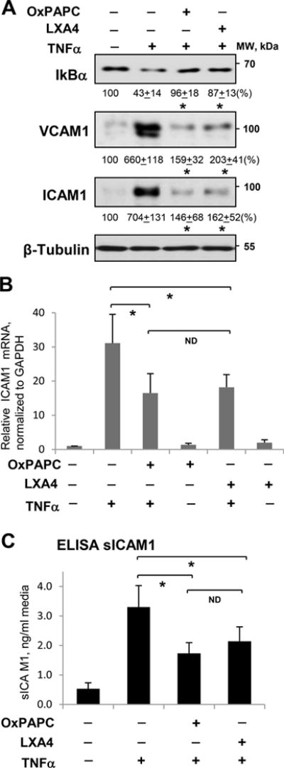
Figure 3. Effect of OxPAPC and LXA4 on TNFα-induced inflammatory activation of human pulmonary EC.

Cells were treated with TNFα (20 ng/ml, 6 hrs) alone or pretreated with OxPAPC (15 μg/ml) or LXA4 (100 nM) for 30 min. A - IκBα degradation, ICAM1 and VCAM1 expression were analyzed by Western blotting. Probing for β-tubulin was used as a normalization control. Numerical data depict results of quantitative densitometry; n=4; p<0.05 vs. TNFα alone. B - Expression of ICAM1 mRNA in control and stimulated HPAEC was evaluated by qRT-PCR; n=3, *P < 0.05 vs. TNFα alone. C - The level of soluble ICAM1 (sICAM1) in EC conditioned medium after stimulations was measured using ELISA assay; n=5, *P < 0.05 vs. TNFα alone.
