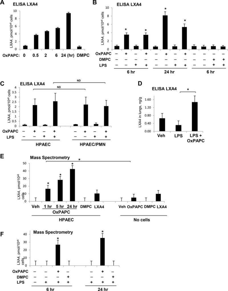
Figure 6. Analysis of OxPAPC-induced LXA4 generation.

EC were treated with OxPAPC (15 μg/ml), DMPC (15 μg/ml), OxPAPC + LPS (200 ng/ml), or DMPC + LPS. A – ELISA assay: time course of LXA4 generation by EC treated with OxPAPC, DMPC, or vehicle; n=3, *P < 0.05 vs. vehicle. B - LXA4 generation by EC challenged with LPS (1 hr) and post-treated with OxPAPC (6 or 24 hrs), DMPC (6 hrs), or vehicle; n=4, *P < 0.05 vs. vehicle. C - Generation of LXA4 by EC and in EC-PMN co-culture. Similar levels of LXA4 were detected in EC cultured alone and in EC co-cultured with PMN; n=4, ND – no difference. D - Increased LXA4 levels in lung tissue of LPS-challenged mice after 5-hrs post-treatment with OxPAPC; n=3, *P < 0.05. E - Mass spectrometry analysis of LXA4 generation by cultured EC. Cells were treated with OxPAPC (1–24 hrs), DMPC (1 hr) or LXA4 (1 hr) as a positive control. Culture medium with addition of OxPAPC, DMPC, or LXA4 without exposure to the cells was used as an additional control. F - Mass spectrometry analysis of LXA4 in the conditioned medium of EC stimulated with LPS, LPS+OxPAPC, or LPS+DMPC. Cells were incubated with agonists for 6 or 24 hrs. LXA4 was detected in the conditioned medium; n=3, *P < 0.05 vs. LPS alone.
