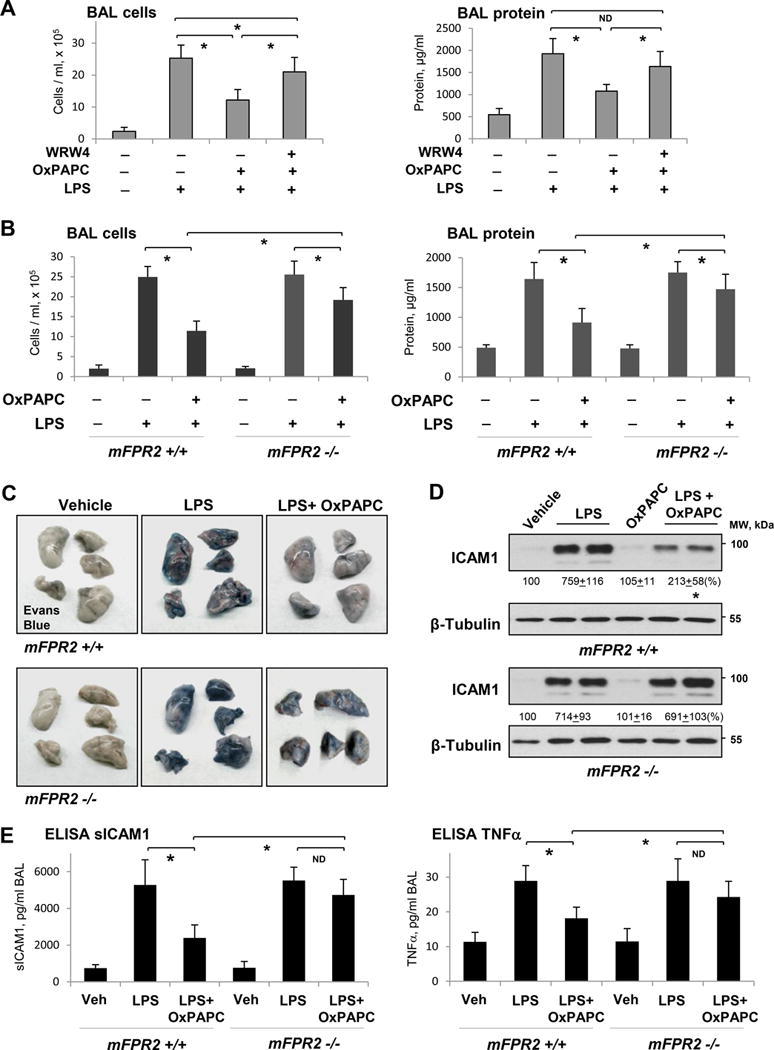
Figure 8. Role of FPR signaling in protective effects of OxPAPC in the model of LPS-induced lung injury.

A - Intravenous injection of OxPAPC (1.5 mg/kg) with our without FPR2/ALX inhibitor WRW4 (20 μM, 1 hr prior to OxPAPC) was performed 5 hrs after LPS instillation (0.7 mg/kg, i.t.). BAL cell count and protein content were measured after 24 hrs of LPS challenge; n=5, *P < 0.05. B - BAL cell count and protein content in mFPR−/− mice and matching controls treated with LPS and LPS+OxPAPC; n=4, *P < 0.05. C - Evans Blue accumulation in the lung parenchyma. D - ICAM1 expression in lung tissue samples of control and mFPR-/- mice after LPS or LPS+OxPAPC challenge. E - ELISA assay of TNFα and sICAM1 levels in BAL samples; n=4, *P < 0.05.
