
Figure 1. Overall structure of MmTPC1.
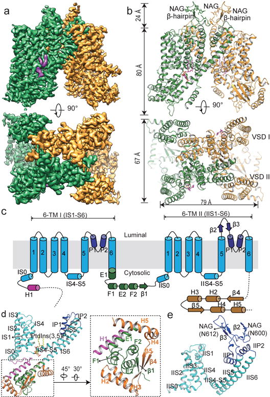
a, 3D reconstruction of PtdIns(3,5)P2-bound (purple density) MmTPC1 dimer with each subunit in individual color. b, Cartoon representation of MmTPC1 in the same orientations as the EM maps in a. N-acetylglucosamine (NAG) molecules and PtdIns(3,5)P2 are shown as sticks. c, Topology and domain arrangement of MmTPC1 subunit. d, Structure of the 6-TM I and the soluble domain with individual element colored as that in c. Inset: zoomed-in view of the cytosolic soluble domain. e, Structure of the 6-TM II.