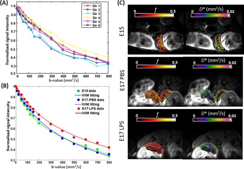
Fig. 2.
IVIM analysis of placental capillary perfusion. (A) Diffusion-weighted signals acquired from a representative E17-PBS placenta at 16 b-values and six diffusion directions. (B) Measured and fitted diffusion-weighted signals in representative E15, E17 PBS, E17 LPS placentas, averaged over six diffusion directions. (C) Maps of the pseudo-diffusion fraction (f) and pseudo-diffusion coefficients (D*) fitted from the IVIM model and mapped onto non-diffusion-weighted images, in an E15, an E17 PBS-treated, and an E17 LPS-treated mouse.

