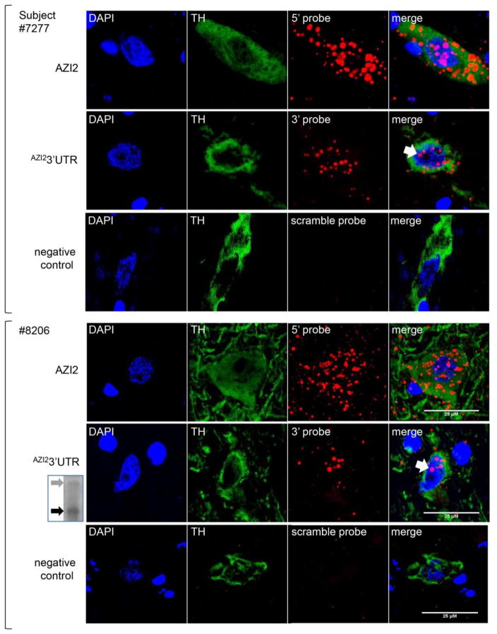
Figure 3. Nuclear localization of 3′UTR expression in human post mortem nigral dopamine neurons from two individuals (upper three rows from #7277, a 58 year old male and lower three rows from #8206, a 93 year old female) based on confocal images from RNAscope analysis.
The consistent findings, despite different demographics, suggests the generalizability of these findings. Three up-to-down rows represent three RNA probes, 5′ probe for AZI2, 3′ probe for 3′UTR and scrambled probe for negative control; four left-to-right columns of images are DAPI for nucleus, TH (tyrosine hydroxylase protein immunostaining) for labeling DA neurons, RNA probing, and merging all three. White arrow, nuclear expression of 3′UTR by 3′ probe; Scale bars, 25 μm. Insert on the lower left: Northern blot for enriched expression of AZI23′UTR (black arrow), compared to AZI2 (gray arrow), in the same female (see supplementary data for an enlarged view).

