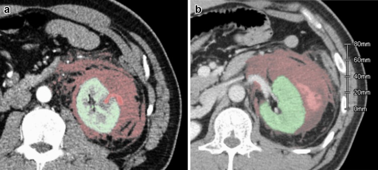
Fig. 2.
Computed tomography (CT) image with contrast to examine the cause of sudden flank pain 7 days after the procedure. As shown in an arterial phase, expansion of the hematoma was explained by the extravasation protruding outward from the renal parenchyma (a). At the equilibrium phase, the contrast was viewed within the hematoma, representing active bleeding (b). A hematoma volume of 955 mL, shown as the red-colored area, was measured using reconstructed CT data

