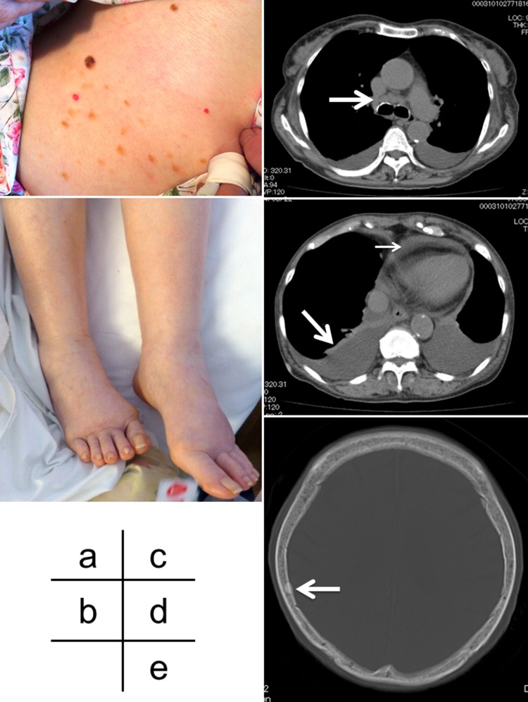
Fig. 1.
a Multiple raised erythematous lesions 5 mm in size on the trunk. b Edema of the bilateral lower extremities. c Computed tomography shows an enlarged mediastinal lymph node measuring 15 mm in size (arrow). d Computed tomography showed bilateral pleural effusions (large arrow) and slight accumulation of pericardial fluid (small arrow). e Computed tomography displays a small sclerotic lesion measuring 10 mm (arrow)

