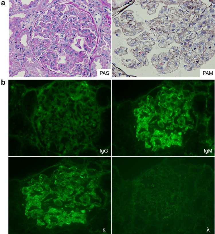
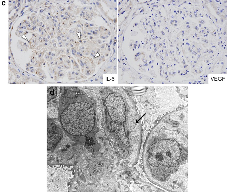
Fig. 2.
Renal biopsy specimen. a There are diffuse lobular endocapillary proliferative changes with endothelial cell swelling, as well as infiltration of monocytes and neutrophils. b Immunofluorescence demonstrates positive staining of the glomerular capillaries and mesangium for IgM and κ light chain. c Immunohistochemical staining shows that podocytes are weakly positive for IL-6 (arrow head), but VEGF is negative. IL interleukin, VEGF vascular endothelial growth factor. d Electron microscopy reveals endothelial cell swelling and edematous expansion of the subendothelial space (arrow) in the GBM with no electron-dense deposits. GBM glomerular basement membrane


