Fig. 6.
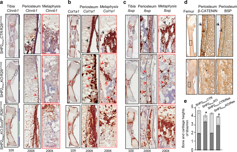
SHP2 deficiency compromises osteogenic differentiation of OCPs and skeletal ossification, and can be rescued by haploinsufficiency of Sox9. a–c Frozen sections from 1.5-day-old mouse tibiae, hybridized to probes for murine Ctnnb1 (a), Col1α1 (b), and Ibsp (c). Enlarged views of the corresponding color-boxed areas are presented on the right. Note the reduced abundance of Ctnnb1, Col1α1, and Ibsp in cells within the periosteum and spongy bone in SHP2Prrx1KO mice (middle row), compared with SHP2Prrx1CTR mice (top row). These defects and defective ossification were partially rescued by Sox9 haploinsufficiency in SHP2Prrx1KO;Sox9fl/+;R26mTmG mice (bottom row). d Images of femurs from 7-day-old SHP2Prrx1CTR and SHP2Prrx1KO mice, immunostained with antibodies against β-CATENIN and bone sialoprotein (BSP). Note the reduction of β-CATENIN and BSP abundance in periosteal areas (*) of SHP2Prrx1KO mice, compared with SHP2Prrx1CTR controls (arrows). Arrowheads indicate calcified cortical bone in SHP2Prrx1CTR mice (a–d, n = 3). e Bar graphs demonstrate the heights of epiphyseal cartilage (dark gray) and endochondral bone (light gray) of tibia from P0.5 neonates. Note that removal of one allele of Sox9 significantly increased the length of endochondral bone in SHP2OCPKORes mice, compared to controls (n = 3, *#P < 0.05, 2-way ANOVA).

