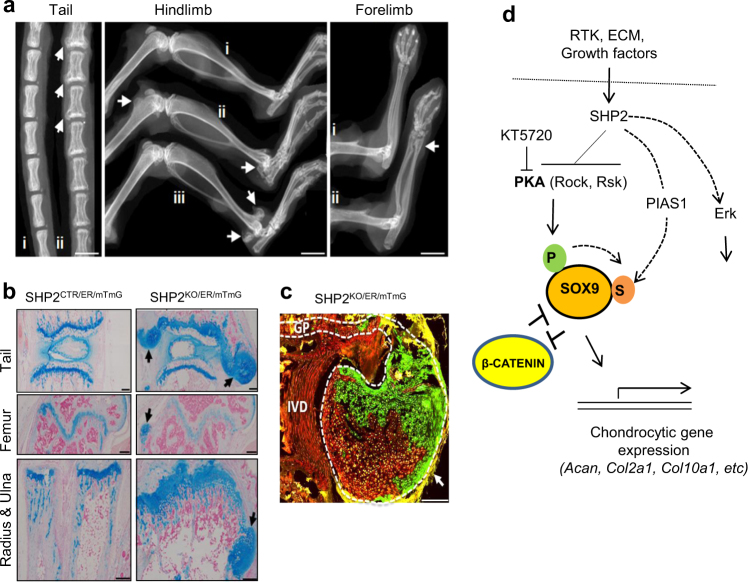
Fig. 7.
Mosaic Ptpn11 deletion at E13.5 in Prrx1-CreERt2-expressing OCPs leads to osteochondromas and enchondromas. a High resolution plain radiographs showing the outgrowth of cartilaginous lesions (arrows) on the tail vertebrae, distal femur, distal tibia, radius and ulna (n = 14). i: SHP2CTR/ER/mTmG; ii, iii:SHP2LOH/ER/mTmG. b Images of tail vertebrae, distal femur, and ulna sections, stained with Alcian blue and nuclear fast red, demonstrating cartilaginous lesions (arrows) in SHP2KO/ER/mTmG mice, compared with SHP2CTR/ER/mTmG mice. Note that there is no clear separation between the growth plates of the radius and ulna in SHP2LOH/ER/mTmG mice. c Merged fluorescent microscopic images showing cells in which the Rosa26mTmG reporter allele has (green fluorescence) or has not (red fluorescence) been recombined by Cre recombinase in a SHP2LOH/ER/mTmG mouse. A large cartilage outgrowth (arrow) adjacent to the intervertebral disc (IVD) and tail vertebral growth plate (GP) is shown. The GP and cartilage outgrowth are outlined (white dashed line). Note that green and red chondrocytes are observed in the outgrowth. Scale bars in a represent 4 mm; Scale bars in b and c represent 250 μm (For the studies in b and c, n = 3 for each genotype). d Proposed model of how SHP2 regulates chondrogenesis and endochondral bone formation by altering the expression of the transcription factors SOX9 and β-CATENIN. P, phosphorylation; S, SUMOylation.

