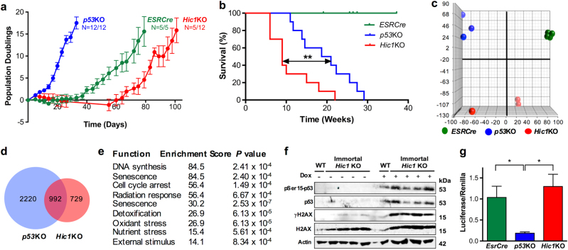
Fig. 2. Immortalization of MEFs lacking Hic1 results in a phenotype distinct to MEFs lacking p53.
a Growth of MEFs (shown as cumulative population doublings) following tamoxifen treatment using the 3T3 protocol [65]. Data shown as mean+SEM. Sample size was determined by the number of available immortalized MEF lines. Cell lines were checked for Mycoplasma and genotype every 6 months. b Kaplan–Meier survival analysis of athymic nude mice injected with 1 × 10⁶ immortalized MEFs with the genotypes indicated. **P < 0.005, log-rank analysis. 1 × 10⁶ MEFs were resuspended in 50 µl media+50 µl Matrigel and injected subcutaneously in the right flank and observed for 26 weeks, until the tumor reached 800 mm3 measured by an observed blinded to the MEF genotype. c Principal component (PC) analysis of gene expression in immortalized MEFs generated from embryos with the genotypes indicated compared to control MEFs. Detailed bioinformatic methods are described in Supplementary Information. Array data are available through GEO, GSE104394. d A Venn diagram depicting differentially expressed genes in the immortalized MEF lines shown in Fig. 1c when compared with control MEFs. e Gene ontology analysis of differentially expressed genes when comparing immortalized p53KO vs. Hic1KO MEFs. The analysis was performed by an observer with no a priori knowledge of the cellular phenotype. f Western blot analysis of lysates from wild type (WT) or Hic1 KO MEFs showing the expression of p53 phosphorylated at serine 15 (pSer15-p53, Cell Signaling Technology, Danvers, MA, USA, #9284S), p53 (Santa Cruz Biotechnology, Dallas, TX, USA, sc-6243), phosphorylated-γH2AX (γH2AX, Novus Biologicals, Littleton, CO, USA #NB100-74435), total H2AX (Cell Signaling Technology, 2595 S) and Actin [66, 67]. Cells were treated with vehicle or doxorubicin (Dox; 1 μM, 6 h). g Activity of a p53-responsive luciferase reporter (Qiagen, Hilden, Germany, #CCS-004L) in MEFs 24 h after treatment with doxorubin, 1 μM, for 6 h. n = 4 indepdent cell lines performed, each performed in triplicate, mean + SEM, *P < 0.05

