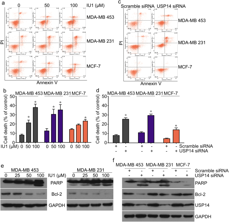
Fig. 6.
USP14 inhibition or silencing induces apoptosis in AR+/ER– breast cancer cells. The indicated breast cancer cell lines treated with IU1 for 72 h were collected. Flow cytometry analysis with annexinV-FITC/PI staining was used to calculate the apoptotic cells. Representative images (a) and cell death population (b) from three independent replicates are shown. Mean ± S.D. (n = 3). *P < 0.05 vs. each vehicle control. The indicated breast cancer cells treated with USP14 siRNA or control siRNA for 72 h were collected. Flow cytometry analysis with annexinV-FITC/PI staining was used to calculate the apoptotic cells. Representative images (c) and cell death population (d) are shown. Mean ± S.D. (n = 3). *P < 0.05 vs. each vehicle control. e Protein lysates were collected from the indicated breast cancer cells treated with IU1 for 48 h. Western blot assay was used to detect PARP and Bcl-2 protein expression. f Protein lysates were collected from the indicated breast cancer cells treated with USP14 siRNA or control siRNA for 48 h. Western blot assay was used to detect PARP and Bcl-2 expression. GAPDH was used as an internal control

