Fig. 1.
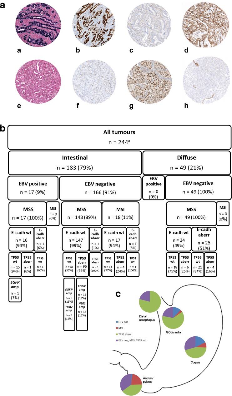
Classification of adenocarcinomas of the stomach, gastro-oesophageal junction and distal oesophagus based on immunohistochemistry and in situ hybridisation. a Examples of EBER in situ hybridisation and MLH1, TP53 and E-cadherin immunohistochemistry in eight oesophagogastric adenocarcinomas. (a) EBV positive, (b) TP53 aberration, (c) MLH1 mutated, (d) E-cadherin wild-type, (e) EBV negative, (f) TP53 wild-type, (g) MLH1 wild-type and (h) E-cadherin aberration. b The classification of the intestinal- and diffuse-type adenocarcinomas of the stomach, gastro-oesophageal junction and distal oesophagus according to the immunohistochemical data and in situ hybridisation. c Distribution of the four molecular subtypes of intestinal-type oesophagogastric adenocarcinomas in different anatomical locations. aImmunohistochemical data available for 238 (EBV, MSI, TP53) or 232 tumours (E-cadherin). bIncluding one tumour (2%) with co-amplification EGFR and HER2. cIncluding five tumours (5%) with co-amplification of EGFR and HER2. EBV Epstein-Barr virus, GOJ gastro-oesophageal junction, MSS microsatellite-stable, MSI microsatellite-instable, wt wild-type, aberr aberration, amp amplification

