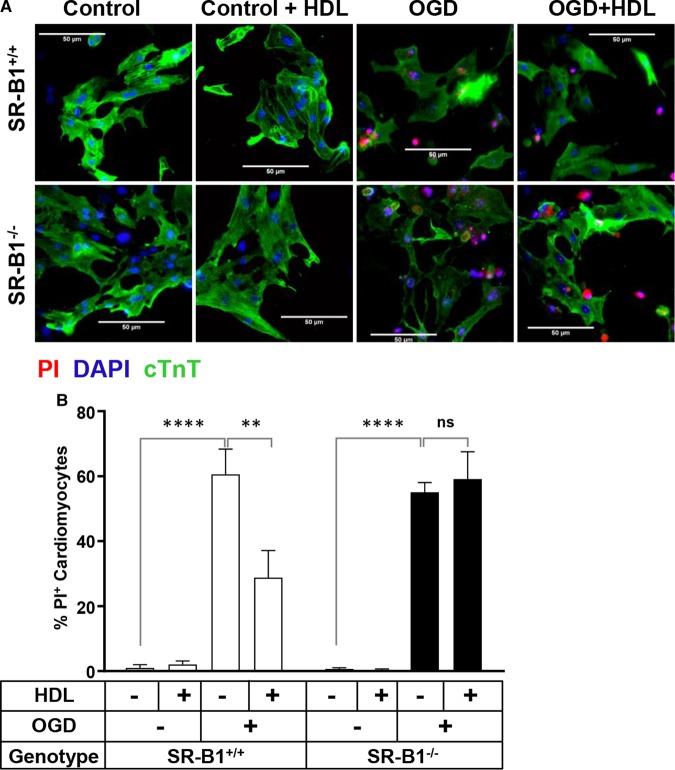
Figure 3. SR-B1 is required for HDL-mediated protection of NMCM against OGD-induced necrosis.
(A) Representative images and (B) quantification of PI staining in SR-B1+/+ and SR-B1−/− NMCM treated for 30 min with or without HDL (100 μg/ml) prior to exposure to OGD for 4 h. Control cells were maintained in normal media under normoxic conditions. In (A), cells were stained prior to fixation with PI (red) or, after fixation, immunostained for cTnT (green) and stained with DAPI (blue). Scale bars = 50 μm. (B) Data represent means ± SEM for n = 3 replicates. ****P < 0.0001; **P < 0.01; ns, not statistically significant (P > 0.99) by one-way ANOVA.

