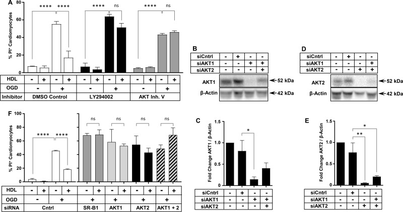
Figure 6. PI3K, AKT1, and AKT2 are required for HDL-mediated protection of HIVCM against OGD-induced necrosis.
(A) Quantification of OGD-induced PI staining in HIVCM treated with or without HDL (100 μg/ml) and inhibitors of PI3K (10 µM LY294002) or AKT (3 µM AKT Inh. V) prior to 4 h of OGD. Control cells were treated with an equal volume of DMSO (0.1%). (B) Representative immunoblots of AKT1 and β-actin (loading control) and (C) quantification of fold change in AKT1/β-actin protein levels in HIVCMs transfected with the indicated siRNAs. (D) Representative immunoblots of AKT2 and β-actin (loading control) and (E) quantification of fold change in AKT2/β-actin protein levels in HIVCMs transfected with the indicated siRNAs. (F) Quantification of OGD-induced necrosis (% PI-positive cells) in HIVCMs transfected with the indicated siRNAs and treated with or without HDL (100 μg protein/ml) for 30 min prior to 4 h of OGD, as indicated. Data in all panels represent means ± SEM (n = 3 replicates). ****P < 0.0001; **P = 0.009; *P < 0.04; ns, not statistically significant (P > 0.33) by one-way ANOVA.

