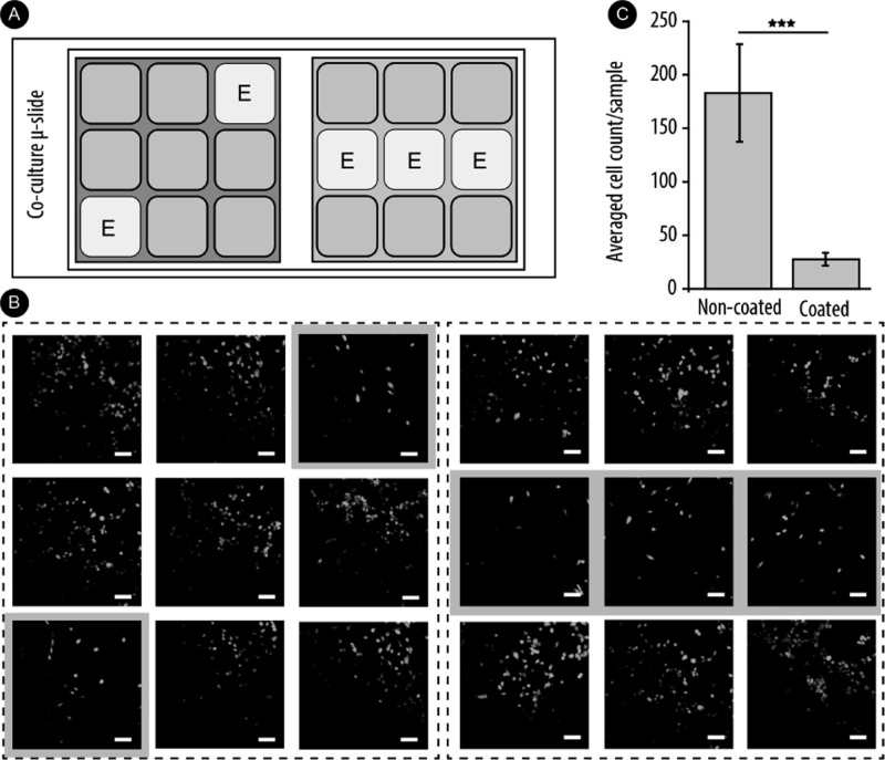
Figure 9.
(A) Schematic representation of coculture μ-slides indicating the multilayered-coated wells. (B) Fluorescence microscopy imaging of myoblast cells. Selected wells were coated with biocatalytic multilayers with 20 mg/L β-Gal for local delivery of NO. Cells were incubated for 48 h in the presence of 100 μM NONOate, replenished at 24 h. Scale bar: 100 μm. (C) Averaged cell count of coated vs noncoated wells. Results are presented as mean ± SD for at least three independent experiments. ***P < 0.001.

